Overview of Retinal Prosthesis and Future Directions
Moon JY, Sather RN, Montezuma SR. Retinal Physician. May 2024;21:9-11. URL: https://www.retinalphysician.com/issues/2024/may/overview-of-retinal-prosthesis-and-future-directions/
Images
-
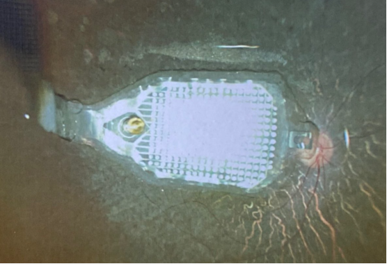
Figure 1 — Fundus photo of IMIE 256 (Golden Eye Bionic / IntelliMicro Medical). Courtesy Mark Humayun MD PhD and Yangyi Yao PhD. Local:
assets/images/retinal-physician-2024/fig1-imie256-fundus.pngSource: https://www.retinalphysician.com/media/ii0jm5os/fundus_0102_3.png -
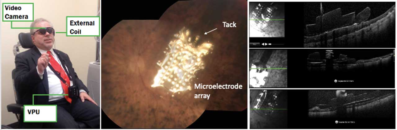
Figure 2 — Argus II epiretinal device (Second Sight Medical Products). External components, fundus photo of 6×10 mm electrode array over macula with tack, and OCT. Patient of Sandra Montezuma MD (published with consent). Local:
assets/images/retinal-physician-2024/fig2-argus-ii.jpgSource: https://www.retinalphysician.com/media/nnyk1gc0/pg10_fig2_may24_web.jpg -
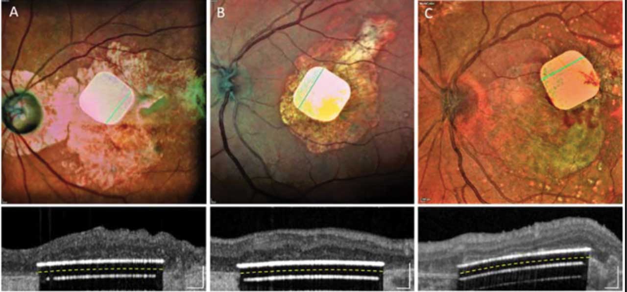
Figure 3 — PRIMA subretinal implant (Pixium Vision). Top: fundus photos of prosthesis in 3 patients with advanced GA. Bottom: OCT placement under macula. Reprinted from Palanker D et al. Ophthalmology 2020;127(8):1097-1104 (open access). Local:
assets/images/retinal-physician-2024/fig3-prima-fundus-oct.jpgSource: https://www.retinalphysician.com/media/y2cbnptn/pg10_fig3_may24_web.jpg
Summary
A 2024 clinical review of 15 retinal and visual prosthesis devices across anatomical placement categories (epiretinal, subretinal, suprachoroidal, optic disc, cortical). Published by University of Minnesota ophthalmology group; co-author Montezuma has past consultancy disclosures to Second Sight and Pixium Vision.
Device table extracted
Epiretinal
| Device | Manufacturer | Stage | CT ID | Notes |
|---|---|---|---|---|
| IMIE 256 | Golden Eye Bionic (US) + IntelliMicro Medical (CN) | Clinical | — | 5 patients, RP end-stage; 256 ch; all improved on all performance measures; 1 AE (electrode movement + low IOP). Xu et al. TVST 2021;10(10):14. |
| EPI-RET 3 | RWTH Aachen University, DE | Clinical (completed, historical) | — | 6 patients; removed at 1 month (ethics committee). No AEs. VA: light perception to hand motion. Menzel-Severing et al. Eye 2012;26(4):501-509. |
| NR600 | Nano Retina, IL | Terminated | NCT04295304 | 9 patients; trial terminated end-2023; lack of funding. |
| Argus II | Second Sight Medical Products, US | Discontinued | NCT03418116 | FDA-approved 2013; >350 patients; discontinued 2019. |
| Polyretina | Diego Ghezzi lab (EPFL/USI) | Preclinical | — | Foldable, photovoltaic, wide-field epiretinal; activates RGCs at safe irradiance; 2-yr in vitro lifetime; no cytotoxicity. Ferlauto et al. Nat Commun 2018;9:992. |
Subretinal
| Device | Manufacturer | Stage | CT ID | Notes |
|---|---|---|---|---|
| PRIMA | Pixium Vision / Science Corp. | Commercial (EU) | NCT03392324, NCT04676854, NCT03333954 | 38-patient PRIMAvera confirmatory trial; CE mark Jul 2026; see raw/papers/holz-2026-prima-subretinal-photovoltaic.md |
| HARP4k | Iridium Medical Technology, TW | Preclinical | — | Computer simulation confirmed safety (minimal mechanical stress, reduced retinal tear risk). Website: irmedtech.com |
| Boston Retinal Implant | Boston Retinal Implant Project; commercial partners: Bionic Eye Technologies + Visus Technology, US | Preclinical | — | 256+ independently configurable channels; wireless power; titanium case on sclera; thin flexible electrode array only enters eye; subretinal. bostonretinalimplant.org |
Suprachoroidal
| Device | Manufacturer | Stage | Notes |
|---|---|---|---|
| Bionic Eye System Gen 2 | BVT / CERA, AU | Phase 2 completed | 4 patients; safety confirmed; meaningful vision gains on screen-based functional assessments; further modifications underway. |
| Suprachoroidal-transretinal stimulation | Osaka University Graduate School of Medicine, JP | In vivo studies | 3 patients; favorable safety profile; variable functional results; 49-microelectrode array in scleral pocket. Fujikado et al. IOVS 2011;52(7):4726-4733. |
| Phoenix99 | University of Sydney + Bionics Institute, AU | Preclinical | 98 stimulation sites + 1 common electrode; bovine in vivo model; safety confirmed. Eggenberger et al. Biomaterials 2021;279:121191. |
Optic Disc
| Device | Manufacturer | Stage | Notes |
|---|---|---|---|
| AV-DONE | Osaka University Medical School, JP | Clinical (1 patient) | Electrode implanted into optic disc; 1 patient 2009 (phosphenes in 6/7 electrodes); original design dislodged → second-generation 7 stim + 1 ref electrodes with 1-step implantation; tested in rabbit + 1 human volunteer. Sakaguchi et al. J Artif Organs 2009;12(3):206-209. Nishida et al. Brain Stimul 2015;8(3):678-681. |
Cortical
| Device | Manufacturer | Stage | CT ID | Notes |
|---|---|---|---|---|
| Orion | Cortigent (Vivani Medical), US | Active (EFS done, pivotal planning) | — | 60 electrodes on medial occipital surface; 5-yr EFS (6 patients, 3 still participating). |
| Cortical Vision Neuroprosthesis | Biomedical Technologies SL, ES | Clinical | NCT02983370 | Intracortical layer; monkey pre-clinical (visual potential confirmed); 1 human trial (consistent stimulation thresholds). biomedical-technologies.com |
| ICVP | Illinois Institute of Technology, US (Chicago Lighthouse) | Phase 1 | — | Floating microelectrode arrays; wireless; no scalp connectors; 1 patient implanted; phase 1 recruiting. chicagolighthouse.org/icvp |
References (article)
- Ghezzi D. Front Neurosci. 2015;9:290.
- Nowik K et al. J Clin Neurosci. 2020;78:8-19.
- Yanovitch L et al. bioRxiv. doi:10.1101/2022.09.14.507901
- Xu H et al. Transl Vis Sci Technol. 2021;10(10):14. doi:10.1167/tvst.10.10.14
- Ferlauto L et al. Nat Commun. 2018;9(1):992. doi:10.1038/s41467-018-03386-7
- Menzel-Severing J et al. Eye (Lond). 2012;26(4):501-509. doi:10.1038/eye.2012.35
- Ostad-Ahmadi Z et al. Int J Ophthalmol. 2021;14(2):310-316.
- Loewenstein JI et al. Arch Ophthalmol. 2004;122(4):587-596.
- Ramirez KA et al. Int J Retin Vitr. 2023;9(1):73.
- NCT03392324 (PRIMA US-FS)
- Iridium Medical Technology: irmedtech.com
- Boston Retinal Implant: bostonretinalimplant.org
- Muqit M et al. medRxiv 2023. doi:10.1101/2023.11.12.23298227
- Petoe MA et al. Transl Vis Sci Technol. 2021;10(10):12.
- Eggenberger SC et al. Biomaterials. 2021;279:121191.
- Fujikado T et al. IOVS. 2011;52(7):4726-4733.
- Bloch E et al. Ther Adv Ophthalmol. 2019;11:2515841418817501.
- Nishida K et al. Brain Stimul. 2015;8(3):678-681.
- Sakaguchi H et al. J Artif Organs. 2009;12(3):206-209. 20-24. Wang V, Harris M, Vivani PR, biomedical-technologies.com, chicagolighthouse.org/icvp 25-28. Dagnelie G, Hosseini Shabanan S, Drag S, Wang C (future directions references)